Hip dysplasia
The hip joint is composed of the ball and the socket. The development of hip dysplasia is determined by an interaction of genetic and environmental factors, though there is a complicated pattern of inheritance for this disorder, with multiple genes involved. Hip dysplasia is the failure of the hip joints to develop normally (known as malformation), gradually deteriorating and leading to loss of function of the hip joints.
Hip dysplasia is one of the most common skeletal diseases seen in dogs. Gender does not seem to be a factor, but some breeds are more likely to have the genetic predisposition for hip dysplasia than other breeds. Large and giant breeds are most commonly affected, including the Great Dane, Saint Bernard, Labrador Retriever, and German Shepherd. Rarely, small breed dogs can also be affected, but are less likely to show clinical signs.
Hip dysplasia often begins while a dog is still young and physically immature. Early onset usually develops after four months of age. There are also cases of later onset, where hip dysplasia develops later due to osteoarthritis, a form of joint inflammation (arthritis) that is characterized by chronic deterioration, or degeneration of the joint cartilage.
Source: http://www.petmd.com/dog/conditions/musculoskeletal/c_dg_hip_dysplasia
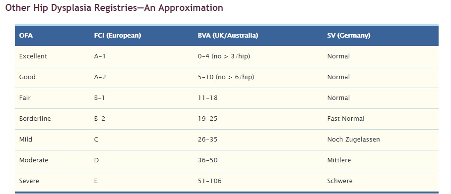
Read more: http://www.offa.org/hd_treatment.html
Elbow dysplasia
Elbow dysplasia is a condition caused by the abnormal growth of cells, tissue, or bone. The condition is characterized by a series of four developmental abnormalities that lead to malformation and degeneration of the elbow joint. It is the most common cause of elbow pain and lameness, and one of the most common causes of forelimb lameness in large and giant-breed dogs. Labrador retrievers, Rottweilers, Golden retrievers, German shepherd dogs, Bernese mountain dogs, chow chows, bearded collies, and Newfoundland breeds are the most commonly affected. The age for onset of clinical signs is typically four to ten months, with diagnosis generally being made around 4 to 18 months.
One type of the condition is more likely to affect males than females: when the bone fragment is located at the inner surface of the upper ulna. The ulna is one of the bones of the foreleg, just below the elbow joint. Otherwise, there are no known gender differences.
Source: http://www.petmd.com/dog/conditions/musculoskeletal/c_dg_elbow_dysplasia
Read more: http://www.offa.org/pdf/elbowarticle.pdf